What is Digital Pathology?
Pathology is the diagnosis and analysis through the examination of tissue from the human body, which is traditionally fixed on glass slides and reviewed under a microscope. Over the past two decades years, technology has played a pivotal role in changing the landscape of several industries by digitizing traditional analog processes, and pathology hasnít been left out. Digital pathology is gained popularity as increasing numbers of laboratories started to adopt new technologies.
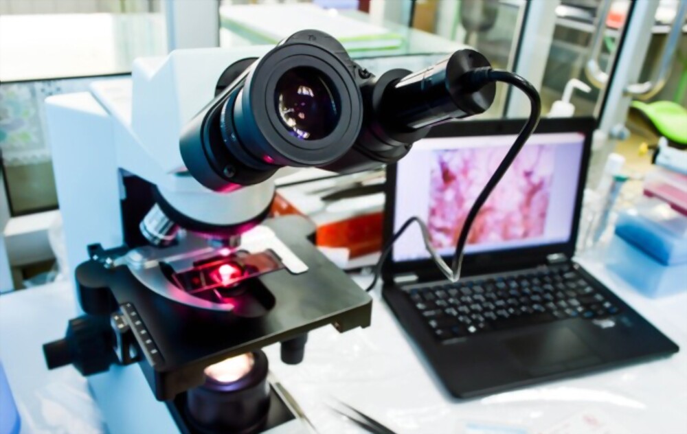
Digital pathology, as a term, can have broad or narrow meanings. It can be defined broadly as the usage, including display and analysis, of digitally acquired images of pathological samples to attain a clinical or research objective. Digital pathology can have an array from photo-micrographs through with a microscope?mounted digital camera, to remote viewing of video images, to digital whole-slide imaging (WSI) Ė this includes viewing and interpretation of the digital images by a human being, as well as manipulation, analysis or interpretation of the data by software.
Digital Pathology Market
It is important to be mindful of this broad definition because many think only of WSI technology as digital pathology, which overlooks business cases for other uses of digital images and the growth of the digital pathology market. However, WSI is a digital technology that demand huge organised commitment and investment. It allows the scanning of glass slides entirely, with an output of their digitized reproduction that are of diagnostic quality. As the WSI industry continues to advance, the US Food and Drug Administration has approved two of the commercially available scanners, which has paved the way for the usage of WSI as a primary diagnostic tool. By the end of 2023, the digital pathology market is estimated to be valued at USD 726.2 Million.
Aging demographics along with rising incidences of cancer and diseases globally created a high demand and increased workload for pathologists. However, as the demand for pathologists keeps on rising, the number of pathologists in the profession has started declining.
Digital pathology can improve quality
Digital pathology can now be indisputably considered the future of the field. The flexibility, accuracy, and potential to deploy ever-improving informatics and image analysis tools make it an appealing option. A digital workflow provides reduced errors in diagnostic pathology. The involvement of WSI scanner in the histology lab has brought in several advantages over standard microscopy with an overall goal to reduce and prevent errors. In the last decade, the world witnessed a revolution in technological advances with an increase in the use of machine learning (ML), deep learning (DL), and artificial intelligence (AI) tools. Their incorporation in healthcare as well as in diagnostic pathology has set a major glide in the workflow. Artificial intelligence is still in its early stages for clinical practice, but offers potential solutions to manage increased workload of pathologists, while at the same time delivers more steady and timely diagnostic care. It will eventually add to the potential of pathologists to review more cases and provide more accurate diagnoses. More recently, pathologists have had better insight in differentiating between benign and malignant prostate tumors, as well as their architectural patterns with the help of deep learning systems.
Pathological diagnosis requires documentation and pictures. AI in diagnostic pathology has helped built tools for rapid search in large image database to look for an imagery data that may have no annotations or labels. There is an often absence of localized annotation of the area of interest, such as tumor, in digital pathology image databases. Annotations are mostly limited to few features like tumor grade, histologic subtype, etc., rather than a comprehensive morphologic assessment. AI technologies are also helping in creating image search engines and helper tools for pathologists which are need-based image retrieval methods.
Conclusion
Shifting completely to digital in pathology isn't without its challenges. There is still much work remaining in the effort to digitize the pathology labs which is not as simple as changing the workflow to digital and incorporate WSI scanners. Unless there is a rudimentary change in how tissue is processed and therefore the workflow is standardized, and therefore the laboratory has achieved a digital workflow, computer-assisted diagnosis and automatic image analysis aren't possible. Several laboratories have overcome a number of these above challenges and have begun a digital journey. As more laboratories adopt digital pathology and digital images are incorporated into the diagnostic pathology workflow there'll be better case management and resulting of cases due to overall improvement within the information management surrounding a patient case. The convenience of availability of cloud computing, powerful processors, and robust infrastructure today has made it possible to make a pixel-pipeline-based workflow that allows for creating AI-based prognostic or diagnostic algorithms.


